Vestibular System · Volume I of 3
Functional Anatomy and Physiology
Trace how labyrinthine sensors, neural circuits, and reflexes convert motion into gaze, balance, and orientation, making vestibular findings predictable from first principles.
Orientation
About this volume
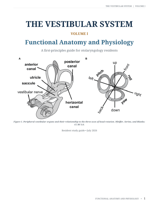
Volume I builds the physiologic model behind the rest of the vestibular series. It follows head motion from the semicircular canals and otolith organs through hair-cell transduction, tonic afferent coding, vestibular nuclei, cerebellar processing, and the reflexes that stabilize gaze, head, body, and perception. Residents work through push-pull canal organization, Ewald’s laws, gravito-inertial ambiguity, reference-frame transformations, velocity storage, multisensory integration, and compensation after loss. The major figures and tables connect physical forces to the eye movements, oscillopsia, and balance consequences seen clinically. Unlike Volume II’s bedside localization and testing, this volume does not organize around disease names. It gives residents a causal model so later examination findings and test disagreements can be explained rather than memorized.
After completing this volume, the resident should be able to
- Trace semicircular-canal and otolith signals from hair-cell transduction through central vestibular pathways.
- Use push-pull organization, Ewald’s laws, and reference frames to predict eye and balance responses.
- Explain how unilateral loss and compensation produce recognizable symptoms and examination findings.
Reader
PDF preview
Preview before loading
The full PDF is loaded only when requested to keep this page fast, especially on mobile.