Facial Trauma · Volume V of 6
Repairing the Facial Envelope
Assess and repair facial soft-tissue wounds as functional injuries, protecting eyelids, lacrimal drainage, facial nerve, parotid duct, free borders, tissue viability, and scar outcomes.
Orientation
About this volume
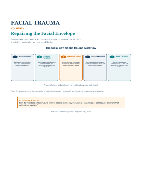
Volume V shifts from the bony framework to the facial soft-tissue envelope, treating wounds as possible disruptions of closure, tear drainage, animation, oral competence, salivary flow, contour, and specialized landmarks. Residents follow a repeatable sequence: examine function before anesthetic, expose the true depth, irrigate and debride conservatively, map critical anatomy, repair in layers, and plan follow-up. The regional chapters address eyelid and canalicular trauma, extratemporal facial nerve injury, parotid and duct trauma, and injuries of the lip, cheek, nose, and ear; later material covers bites, ballistic wounds, scars, and revision. Unlike Volume IV’s mandibular stabilization, this volume centers on structures hidden in the wound itself. Volume VI then integrates skeletal and soft-tissue considerations in panfacial reconstruction.
After completing this volume, the resident should be able to
- Examine facial wounds for hidden injury to function before anesthetic, irrigation, or closure.
- Plan layered repair around eyelid, canalicular, facial-nerve, parotid-duct, muscle, cartilage, and free-border anatomy.
- Match wound contamination, tissue viability, follow-up, and scar biology to safe postoperative planning.
Reader
PDF preview
Preview before loading
The full PDF is loaded only when requested to keep this page fast, especially on mobile.